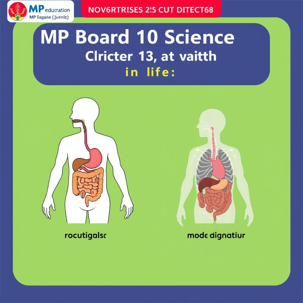
MP Board 10 Science Ch5: Life Processes Notes 2027
MP Board Class 10 Science Chapter 5: Life Processes — Complete Notes, Diagrams & Questions
Welcome to your one-stop study material for MP Board Class 10 Science Chapter 5: Life Processes (जैव प्रक्रम). This chapter covers the fundamental life processes that keep living organisms alive — nutrition, respiration, transportation, and excretion. It’s one of the highest-weightage biology chapters in the MP Board 2027 exam.
📋 Chapter Overview
- Topics Covered: Nutrition (Autotrophic & Heterotrophic), Respiration (Aerobic & Anaerobic), Transportation in Humans & Plants, Excretion in Humans & Plants
- Weightage: 8-10 marks in MP Board 2027 exam
- Key Concepts: Stomata, Human Digestive System, Human Respiratory System, Human Heart & Blood Vessels, Nephron & Excretory System
🌱 1. NUTRITION
1.1 What is Nutrition?
Nutrition is the process by which organisms obtain food and energy for growth, repair, and maintenance. It is of two main types:
A. Autotrophic Nutrition (Auto = self, trophos = nourishment)
- Found in: Plants, algae, and some bacteria (cyanobacteria)
- Process: Photosynthesis — converting light energy into chemical energy
- Equation: 6CO₂ + 12H₂O → C₆H₁₂O₆ + 6O₂ + 6H₂O (in presence of sunlight & chlorophyll)
- Raw materials: CO₂ (from air via stomata), H₂O (from soil via roots), Sunlight (absorbed by chlorophyll), Chlorophyll (green pigment in chloroplasts)
- Site: Chloroplasts in mesophyll cells of leaves
B. Heterotrophic Nutrition (Hetero = other, trophos = nourishment)
Organisms that cannot synthesise their own food and depend on others. Three types:
- Holozoic Nutrition: Ingestion of solid food → digestion → absorption → assimilation → egestion (e.g., humans, amoeba)
- Saprophytic Nutrition: Feeding on dead/decaying organic matter (e.g., fungi, bread mould, bacteria)
- Parasitic Nutrition: Living on or inside a host organism (e.g., tapeworm, lice, Cuscuta)
📐 Diagram: Structure of Stomata
Description: Draw an epidermal peel showing two kidney-shaped guard cells surrounding a pore called the stoma (plural: stomata). Guard cells contain chloroplasts and are connected at their ends. When turgid (water-filled), they curve apart → stoma opens. When flaccid, they come closer → stoma closes. The entire structure is surrounded by ordinary epidermal cells.
Key Labels: Stomatal pore, Guard cells (कवक कोशिकाएँ), Chloroplasts in guard cells, Nucleus, Epidermal cells, Cell wall (thinner outer wall, thicker inner wall)
1.2 Human Digestive System
| Organ | Function | Secretion / Enzyme |
|---|---|---|
| Mouth / Buccal Cavity | Mechanical digestion (chewing), starch breakdown | Saliva → Salivary Amylase (ptyalin) breaks starch → maltose |
| Oesophagus / Food Pipe | Peristalsis — wave-like muscle contractions push food to stomach | No digestion; mucus for lubrication |
| Stomach | Protein digestion, churning, acidic medium | HCl (kills bacteria, activates pepsin), Pepsin (protein → peptones), Mucus (protects stomach lining) |
| Small Intestine (Duodenum + Jejunum + Ileum) | Main site of digestion & absorption | Pancreatic juice (trypsin, lipase, amylase), Bile juice (from liver — emulsifies fats), Intestinal juice (maltase, sucrase, lactase, peptidases) |
| Large Intestine (Colon) | Water absorption, formation of faeces | No digestive enzymes; contains gut bacteria |
| Rectum → Anus | Storage and egestion of undigested food | — |
📐 Diagram: Human Digestive System
Description: Draw the alimentary canal starting from buccal cavity (with tongue and teeth) → pharynx → oesophagus (a long tube behind the trachea) → stomach (J-shaped bag on left side) → small intestine (coiled tube, ~6m long, with finger-like villi) → large intestine (wider, shorter, ~1.5m) → rectum → anus. Show attached glands: Salivary glands (near mouth), Liver (largest gland, right side, stores glycogen, secretes bile stored in gall bladder), Pancreas (below stomach, secretes pancreatic juice containing digestive enzymes and insulin).
Key Labels: Mouth, Oesophagus, Stomach, Liver, Gall Bladder, Pancreas, Small Intestine (Duodenum, Jejunum, Ileum), Large Intestine (Colon), Rectum, Anus, Villi (in inner wall of small intestine)
💨 2. RESPIRATION
2.1 What is Respiration?
Respiration is the process of breaking down glucose to release energy (ATP). It is NOT the same as breathing (which is just the mechanical movement of air).
Types of Respiration
| Feature | Aerobic Respiration | Anaerobic Respiration |
|---|---|---|
| Oxygen required | Yes | No |
| Site | Mitochondria (after glycolysis in cytoplasm) | Cytoplasm |
| Products | CO₂ + H₂O + 38 ATP (net 36-38 ATP) | Ethanol + CO₂ + 2 ATP (in yeast) Lactic acid + 2 ATP (in muscles) |
| Equation | C₆H₁₂O₆ + 6O₂ → 6CO₂ + 6H₂O + Energy | C₆H₁₂O₆ → 2C₂H₅OH + 2CO₂ + Energy (yeast) C₆H₁₂O₆ → 2C₃H₆O₃ + Energy (muscles) |
| Occurs in | Most organisms (humans, plants, animals) | Yeast, bacteria, muscle cells during heavy exercise |
2.2 Human Respiratory System
Pathway of air: Nostrils (Nasal cavity → hair + mucus filter air) → Pharynx (common passage for air & food) → Larynx (voice box) → Trachea (windpipe, supported by C-shaped cartilage rings to prevent collapse) → Bronchi (left & right branches) → Bronchioles (smaller branches) → Alveoli (air sacs — site of gas exchange)
Mechanism of Breathing:
- Inhalation: Diaphragm contracts (flattens) + External intercostal muscles contract → Rib cage moves upwards & outwards → Thoracic cavity volume increases → Lung pressure decreases (below atmospheric) → Air rushes in
- Exhalation: Diaphragm relaxes (domed shape) + Internal intercostal muscles contract → Rib cage moves downwards & inwards → Thoracic cavity volume decreases → Lung pressure increases (above atmospheric) → Air rushes out
📐 Diagram: Human Respiratory System
Description: Draw from top: Nasal cavity → Pharynx → Larynx (Adam’s apple region, containing epiglottis — a flap that prevents food from entering the windpipe) → Trachea (tube with C-shaped cartilage rings) → branching into left and right Bronchi → smaller Bronchioles ending in clusters of balloon-like Alveoli (covered by blood capillaries). Below the lungs, show the dome-shaped Diaphragm.
Key Labels: Nostrils, Nasal Cavity, Pharynx, Larynx, Trachea, Bronchi, Bronchioles, Alveoli, Lungs, Diaphragm, Rib cage, Blood capillaries around alveoli
🫀 3. TRANSPORTATION
3.1 Transportation in Humans — Circulatory System
The Human Heart
The heart is a muscular organ that pumps blood throughout the body. It has 4 chambers:
| Chamber | Receives blood from | Pumps blood to |
|---|---|---|
| Right Atrium (RA) | Superior & Inferior Vena Cava (deoxygenated blood from body) | Right Ventricle (via tricuspid valve) |
| Right Ventricle (RV) | Right Atrium | Lungs (via pulmonary artery) — has pulmonary semilunar valve |
| Left Atrium (LA) | Pulmonary Veins (oxygenated blood from lungs) | Left Ventricle (via bicuspid/mitral valve) |
| Left Ventricle (LV) | Left Atrium | Whole body (via aorta) — thickest wall, has aortic semilunar valve |
Blood Flow Path: Body → Vena Cava → RA → Tricuspid Valve → RV → Pulmonary Valve → Pulmonary Artery → Lungs → Pulmonary Vein → LA → Mitral Valve → LV → Aortic Valve → Aorta → Body
📐 Diagram: Cross-Section of Human Heart
Description: Draw an upside-down pear-shaped organ. Show 4 chambers with a septum (thick muscular wall) dividing left and right halves. Mark Superior Vena Cava (entering RA from top), Inferior Vena Cava (entering RA from bottom), Pulmonary Artery (from RV going to lungs — carries deoxygenated blood — show it as blue), Pulmonary Veins (entering LA from lungs — red), Aorta (from LV — largest artery). Show valves: Tricuspid (RA→RV), Bicuspid/Mitral (LA→LV), and semilunar valves at pulmonary artery and aorta openings.
3.2 Blood Vessels
| Feature | Arteries | Veins |
|---|---|---|
| Direction | Carry blood away from heart | Carry blood towards heart |
| Wall thickness | Thick, elastic, muscular | Thin, less muscular |
| Valves | Absent | Present (prevent backflow) |
| Blood pressure | High | Low |
| Blood type (most) | Oxygenated (except pulmonary artery) | Deoxygenated (except pulmonary vein) |
| Position | Deep inside body | Near skin surface |
3.3 Transportation in Plants
| System | Xylem | Phloem |
|---|---|---|
| What it transports | Water + Minerals (unidirectional: roots → leaves) | Food (sucrose, amino acids) — bidirectional (leaves → storage organs / growing parts) |
| Direction | Only upwards | Upwards and downwards (source → sink) |
| Cells | Tracheids, Vessels, Xylem fibres, Xylem parenchyma | Sieve tubes (living), Companion cells, Phloem fibres, Phloem parenchyma |
| Driving force | Transpiration pull (evaporation from leaves creates suction) | Translocation (uses ATP energy — osmosis at source creates pressure) |
🧪 4. EXCRETION
4.1 Excretion in Humans
Excretory System Organs:
- Kidneys (2): Bean-shaped organs in the abdominal cavity. Filter blood ~180L/day → produce ~1.5L urine. Each kidney contains ~1 million nephrons.
- Ureters (2): Thin tubes carrying urine from kidneys to urinary bladder
- Urinary Bladder: Stores urine temporarily
- Urethra: Carries urine out of the body
4.2 Structure of Nephron (Functional Unit of Kidney)
Parts of Nephron:
- Bowman’s Capsule + Glomerulus (cluster of blood capillaries) = Malpighian Body / Renal Corpuscle
- Proximal Convoluted Tubule (PCT)
- Loop of Henle (U-shaped, extends into medulla)
- Distal Convoluted Tubule (DCT)
- Collecting Duct (common to many nephrons)
Urine Formation — 3 Steps
- ① Glomerular Filtration: Blood pressure forces water, glucose, salts, urea from glomerulus into Bowman’s capsule. Blood cells & proteins stay in blood (too large to pass).
- ② Reabsorption (PCT + Loop of Henle): Useful substances (glucose, amino acids, most water, salts) are reabsorbed into blood capillaries surrounding the tubule.
- ③ Secretion (DCT + Collecting Duct): Excess ions (H⁺, K⁺, NH₃) are secreted into the filtrate. Final urine contains water + urea + excess salts.
📐 Diagram: Nephron
Description: Draw a long tubule starting with a cup-shaped Bowman’s capsule containing a knot of capillaries called Glomerulus (show afferent arteriole bringing blood in, efferent arteriole taking blood out). The capsule continues as PCT (coiled portion in cortex) → a long U-shaped Loop of Henle (descending limb goes into medulla, ascending limb comes back to cortex) → DCT (coiled portion again in cortex) → Collecting Duct (straight tube descending through medulla). Show blood capillaries (peritubular capillaries and vasa recta) wrapping around the tubule for reabsorption.
Key Labels: Bowman’s Capsule, Glomerulus, Afferent Arteriole, Efferent Arteriole, PCT, Descending Limb of Henle, Ascending Limb of Henle, DCT, Collecting Duct, Peritubular Capillaries
4.3 Excretion in Plants
- Waste products: Oxygen (from photosynthesis), CO₂ (from respiration), excess water (transpiration), salts, resins, latex, gums
- Removal methods:
- Gaseous wastes → through stomata (transpiration)
- Solid wastes → stored in dead cells (bark, old leaves, vacuoles), shed periodically
- Resins & gums → stored in resin canals, old xylem vessels
- Excess salts → removed through leaf fall (senescence)
- Latex → stored in laticiferous vessels (e.g., rubber, opium poppy)
📝 IMPORTANT QUESTIONS (MP Board Exam Focus)
Very Short Answer Questions (1 Mark)
- Name the green pigment present in leaves that absorbs sunlight.
- What is the functional unit of the kidney?
- Which enzyme breaks down starch in the mouth?
- What prevents the backflow of blood in veins?
- Name the cell in which aerobic respiration takes place.
Short Answer Questions (2-3 Marks)
- Differentiate between aerobic and anaerobic respiration (any 4 points).
- Write the difference between arteries and veins.
- Explain the process of photosynthesis with chemical equation.
- What is peristalsis? Where does it occur?
- Draw a labelled diagram of stomata and explain its function.
Long Answer Questions (5 Marks)
- Draw a neat labelled diagram of the human digestive system and describe the function of each part.
- Explain the structure and working of the human heart with the help of a labelled diagram. Trace the path of blood from the body to the lungs and back.
- Describe the structure of a nephron and explain the three steps of urine formation.
- Compare xylem and phloem on the basis of their structure, function, and direction of transport.
- Explain the mechanism of breathing in humans. Draw a labelled diagram of the human respiratory system.
NCERT-Based Important Questions (PYQ Pattern)
- Why is small intestine longer in herbivores than carnivores?
- What is the role of HCl in the stomach?
- Why does the wall of the left ventricle have the thickest muscle?
- What happens when we breathe deeply after running?
- Why do plants not have a specialised excretory system like humans?
🔑 Key Terms to Remember
- Autotrophic Nutrition — स्वपोषी पोषण
- Heterotrophic Nutrition — परपोषी पोषण
- Photosynthesis — प्रकाश-संश्लेषण
- Peristalsis — क्रमाकुंचन (wave-like movement in oesophagus)
- Villi — दीर्घरोम (finger-like projections in small intestine)
- Alveoli — वायुकोश (air sacs in lungs)
- Diaphragm — मध्यपट (muscular sheet below lungs)
- Double Circulation — द्विवह परिसंचरण (blood passes through heart twice per cycle)
- Nephron — वृक्काणु (functional unit of kidney)
- Glomerulus — केशिकागुच्छ (capillary network in Bowman’s capsule)
- Transpiration Pull — वाष्पोत्सर्जन खिंचाव (main force for water rise in xylem)
- Translocation — स्थानान्तरण (movement of food in phloem)
💡 Pro Tips for MP Board 2027
- Diagrams are mandatory! Practise drawing all 5 diagrams: Stomata, Digestive System, Respiratory System, Heart, Nephron
- Label everything — unlabelled diagrams get only partial marks
- Write chemical equations — photosynthesis and respiration equations are frequently asked
- Compare & contrast — questions like “difference between” are very common (artery/vein, aerobic/anaerobic, xylem/phloem)
- Hindi key terms — knowing Hindi equivalents (like those above) can help in Hindi-medium exams
Need more practice? Download the MP Board Class 10 Model Paper 2027 from here and practise Chapter 5 questions.
💪 Keep learning, keep growing! Master Life Processes and you’re one step closer to that 90%+ in Class 10 Science 2027.